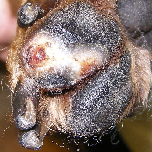


The dog enters the clinic with a paw that had been lacerated several days earlier. The wound is swollen, red and exudating, showing no evidence of granulation tissue. The wound edges are frayed and appear chewed by the dog. The wound is cleaned, SertaSil applied and dressed. Four days later, the wound shows normal healing with formation of granulation tissue. Use of SertaSil is continued and the wound continues to show good healing. Wound closure is achieved in 15 days after start of treatment.
Responsible: Alf Laebro, vet & partner; and Signe L. Dideriksen, vet, Centrum Small Animal Hospital, Denmark.
[toggle border=’none’ title_color=’#0049D1′ title_font_size=’0.75em’ title=’Detailed description:’]A 5 years and 1 month old bitch (Rottweiler mix) enters the clinic limping without touching the ground on the left hind leg. She has some days previously torn her pad and the wound opening is approx. 12 cm2 (3×4 cm). The wound is swollen, red and exudating showing no evidence of granulation tissue. The wound edges are frayed and appear chewed by the dog. Apart from the limp, the bitch seems healthy and happy.
The wound is cleaned with chlorhexidine, air dried, SertaSil is applied and the wound is dressed with a shock resistant and waterproof bandage using Allevyn, Soffban, Vetrap and Tensoplast. An 8 days Amoxicillin treatment is initiated.
The owner is advised to avoid exercise and getting the dressing wet when the dog is outdoors.
The dressing is changed 4 days later. According to the owner, the dog has done very well, and despite having played and chased balls, the wound looks really good. It has pulled together along the edges where granulation tissue is also emerging. The centre of the wound is yellow due to concentration of lymphatic cells. The wound is cleaned with chlorhexidine, air dried, covered with SertaSil and dressed with a shock resistant and waterproof bandage similar to the previous and supplemented with a paw sock.
At the follow-up 2 days later, the dog walks correctly on all four legs without any sign of lameness. The wound looks good. It is virtually no longer exudating, it is beginning to close from the edges toward the centre and is now covered with granulation tissue except for a small yellow spot of lymphoid cells in the centre. The wound is washed, covered with SertaSil and dressed as last time.
At the follow-up 4 days later, the wound again looks good. 4/5 of the wound is now covered by a thin layer of white epithelium, while 1/5 (approx. 1 cm in diameter) is still open, covered with moist surface granulation tissue. Acidity is recognized between the toes because of the dressing, so the hair is cut, the interdigital space is cleaned with chlorhexidine, dried and Fuciderm (fusidic acid + betamethasone) is applied. The original wound is washed, covered with SertaSil and dressed as before.
At the follow-up 2 days later, the remaining part of the wound is also covered with a thin layer of epithelium. There is obvious contraction from the edges and epithelial tissue along the periphery has matured and become stronger and more resilient. Use of SertaSil is discontinued and the paw is simply dressed as previously.
2 days later at the follow-up, the epithelium has matured and become thicker and denser. The paw is cleaned with chlorhexidine and the owner is advised that the dog must wear a paw sock when outdoors but not indoors in order to allow access of air to the toes.
The owner is telephoned 6 days later. Everything is in perfect order. The dog is not wearing a sock and has shown no interest in the injury for the last 5 days. The wound is completely healed and the only trace left is a light discoloration partly due scar tissue.
The wound had closed in 15 days.[/toggle]