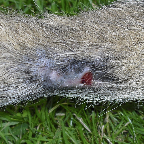
A dog had a 1.4 cm dia. atheroma surgically removed from tail. 10 days later stitches were removed, but the OP site was lightly infected. The owners were instructed to wash with chlorhexidine shampoo and dress the wound daily.
5 days later, the OP wound showed light superficial necrosis. It was cleaned, covered with SertaSil and dressed. The dog had also chewed the tip of the tail and it was warm, oedematous and developing wounds. It was cleaned, but not covered with SertaSil. Baytril (Enrofloxacin) treatment was initiated and Acepromazin was prescribed for 3 nights to avoid further biting.
2 days later, the OP wound had decreased in size and showed obvious improvement. The tip of the tail was still thickened and revealed a distinct festering smell. Both wounds were cleaned, covered with SertaSil and dressed.
4 days later both the tip and the OP-wound showed clear improvement. The wounds were cleaned and covered with SertaSil. The owner was instructed to apply SertaSil on both wounds every other day for a week. At follow-up one week later, both wounds were completely healed.
Responsible: Alf Laebro, vet & partner; and Signe L. Dideriksen, vet, Centrum Small Animal Hospital, Denmark.
[toggle border=’none’ title_color=’#0049D1′ title_font_size=’0.75em’ title=’Detailed description:’]A 3 years and 8 months old Alsatian male dog has had an atheroma of approx. 1.4 cm in diameter surgically removed from the middle of the tail dexter ventrally.
Upon removal of the stitches 10 days after surgery, the OP site was lightly infected. Owners were instructed to wash with chlorhexidine shampoo and dress the wound daily; they were also advised for the dog to wear a cone or otherwise to prevent the dog from licking / biting the area.
At the control visit 5 days later, the wound showed good healing at the bottom of the wound bed but it was accompanied by a light superficial necrosis. The dog had bitten and chewed the tip of the tail considerably, and it was now thickened and displaying wounds in the skin. The tip of the tail seemed warm and oedematous. The OP wound was rinsed with chlorhexidine, SertaSil was applied and the wound dressed with gauze compress, VetRap and Tensoplast. The tip of the tail was also washed with chlorhexidine but SertaSil was not used. A 7 day treatment with Baytril (Enrofloxacin) was initiated and Acepromazin for 3 nights was prescribed. The owner was informed of the risk that the tail could need amputation.
At the control visit 2 days later, the OP wound had decreased in size and showed obvious improvement. The tip of the tail was still thickened and revealed a distinct festering smell. Both wounds were cleaned with chlorhexidine, covered with SertaSil and dressed with Melolin, elastic gauze and VetRap. It was only possible to carry out checks and dressing changes 4 days later, where both the tip of the tail and the OP-wound showed very evident improvement.
Again, the wounds were rinsed with chlorhexidine and SertaSil was applied. The owner brought some SertaSil home and was instructed to use it every other day for a week. At the follow-up visit one week later, everything was completely healed without the need for further treatment.[/toggle]


